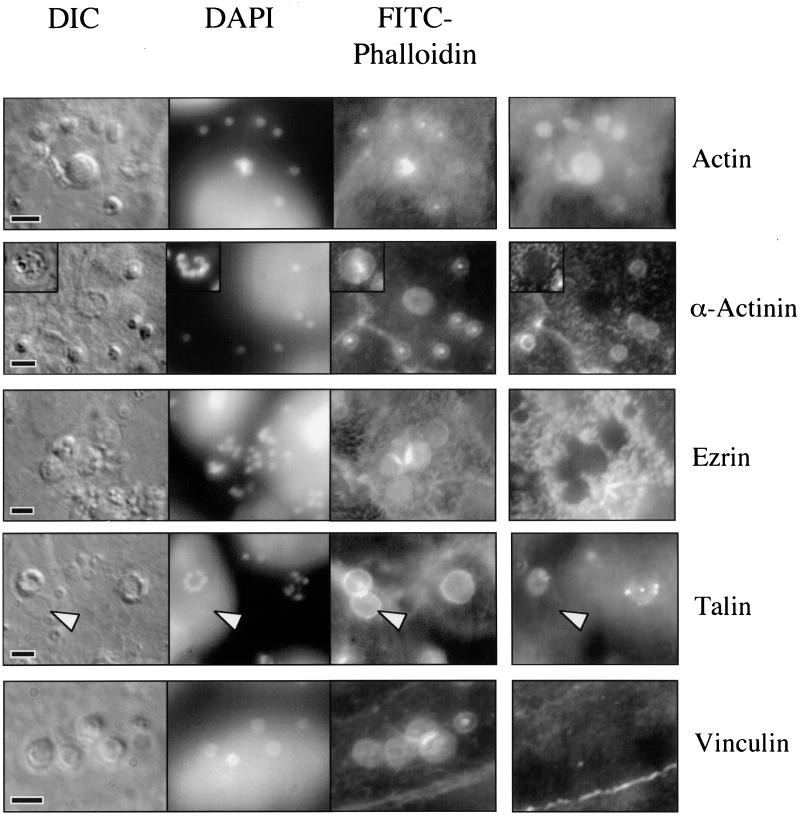
FIG. 6.
Indirect immunofluorescence of actin and actin-associated proteins in a Cryptosporidium-infected cell line. Photomicrographs of a Cryptosporidium-infected cell line reveal DIC images of parasites, parasite nuclei (DAPI), and actin plaques at sites of invasion (FITC-phalloidin). Indirect immunofluorescence using antibodies against actin confirms the presence of actin at each site of invasion in trophozoites (single nuclei) and meronts (center; row 1, column 4). Antibodies against α-actinin (row 2, column 4) reveal the presence of this molecule at sites of developing trophozoites but no staining associated with meronts (insert). Indirect immunofluorescence also shows that ezrin (row 3, column 4) and vinculin (row 5, column 4) are clearly absent from sites of invasion. The linear staining pattern of an adherens junction is shown in the vinculin image (lower right). Although there is some staining by antitalin antibodies (row 4, column 4), the pattern is heterogeneous and is lost when parasites are removed (arrowhead), suggesting that this antibody cross-reacts with the parasite and does not decorate the actin plaque itself. Scale bars = 5 μm.

