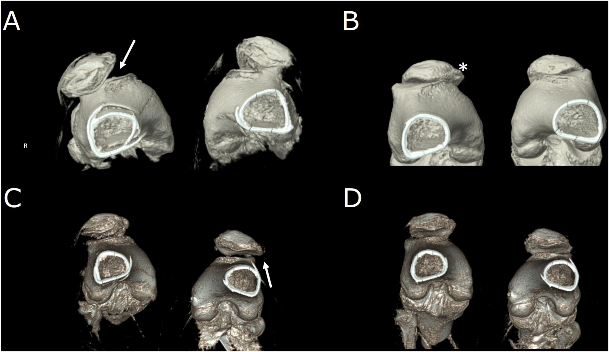
Figure 1.

Pre-and post-operative four-dimensional computed tomography acquisitions of a patient with interval right-sided tibial tubercle osteotomy and medial patellofemoral ligament repair and newly diagnosed left-sided patellar instability. (A) Subluxation in both joints prior to intervention in full extension, worse on the right (arrow). (B) Disappearance of subluxation in full flexion corresponding to the radiographic “J-sign” of maltracking. The presence of osteophyte (*) is consistent with mild-to-moderate secondary osteoarthritis. (C) Full-extension post-operative images show correction of right-sided maltracking with residual left-sided maltracking (arrow). (D) Joints in full flexion post-operatively.
