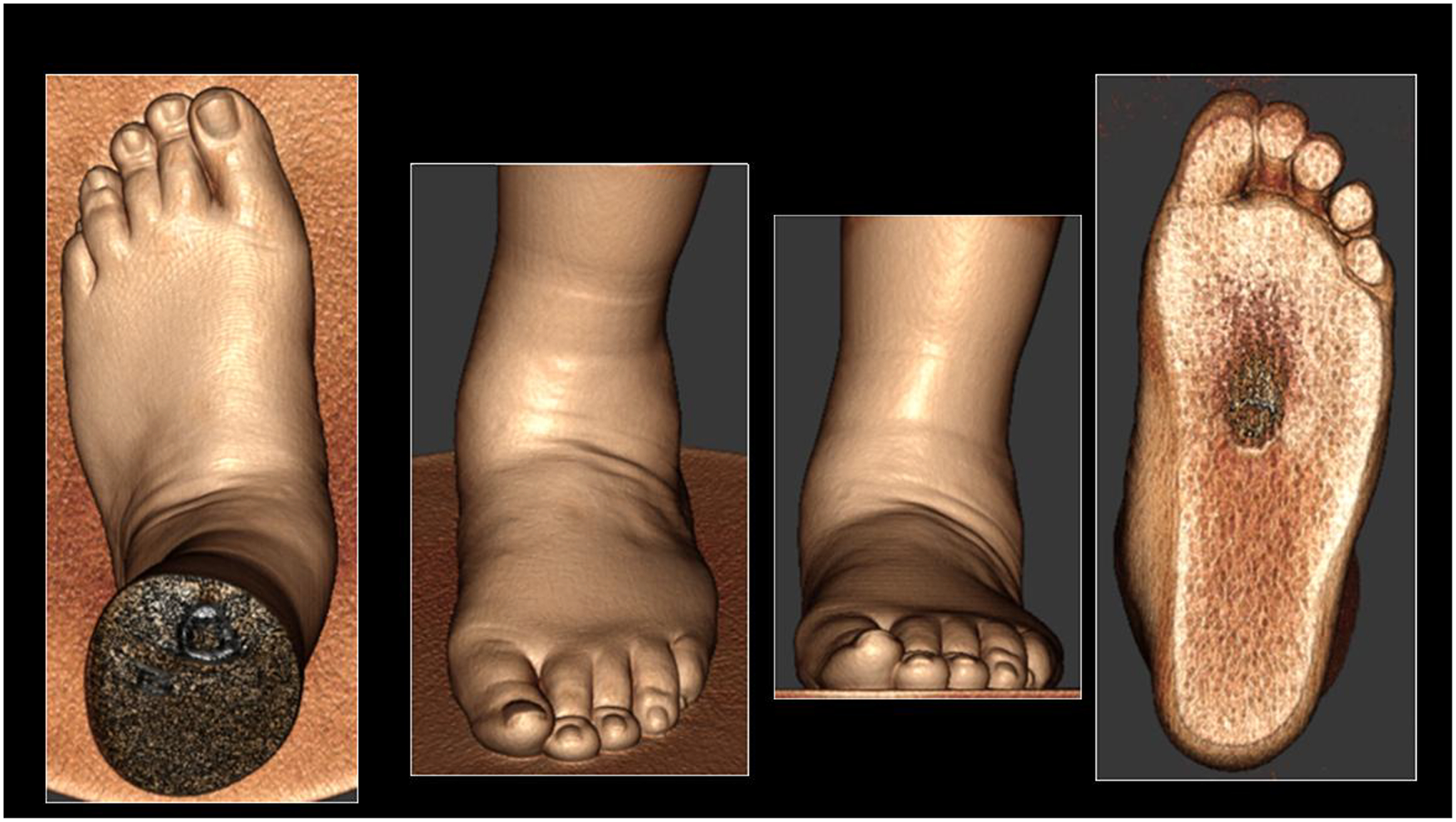
Figure 4:

Three-dimensional rendering of the soft tissue contour of the foot using weight-bearing cone-beam computed tomography of a 56-year-old male suffering from a painful adult acquired flatfoot deformity progressing over eight years. Pain was present around the plantar surface of the heal and sinus tarsi area. Physical exam revealed a flat plantar arch with valgus abduction of the hind-, fore-, and mid-foot regions, tenderness in areas concordant with provided history, limited dorsiflexion, and a hypermobile first ray.
