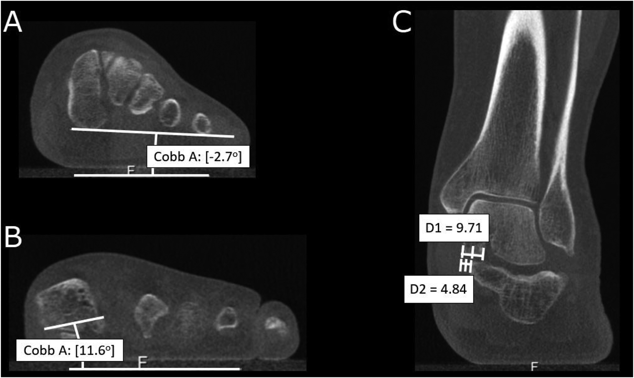
Figure 5.

Measurements from multiplanar reconstructions of weight-bearing cone-beam computed tomography of the patient from figure 4. (A) Measurement of the foot arch angle. (B) First metatarsal base-to-floor angle. (C) ~50% middle facet uncovering, calculated from the division of the measured uncoverage (D2) by the width of the talar middle facet (D1).
