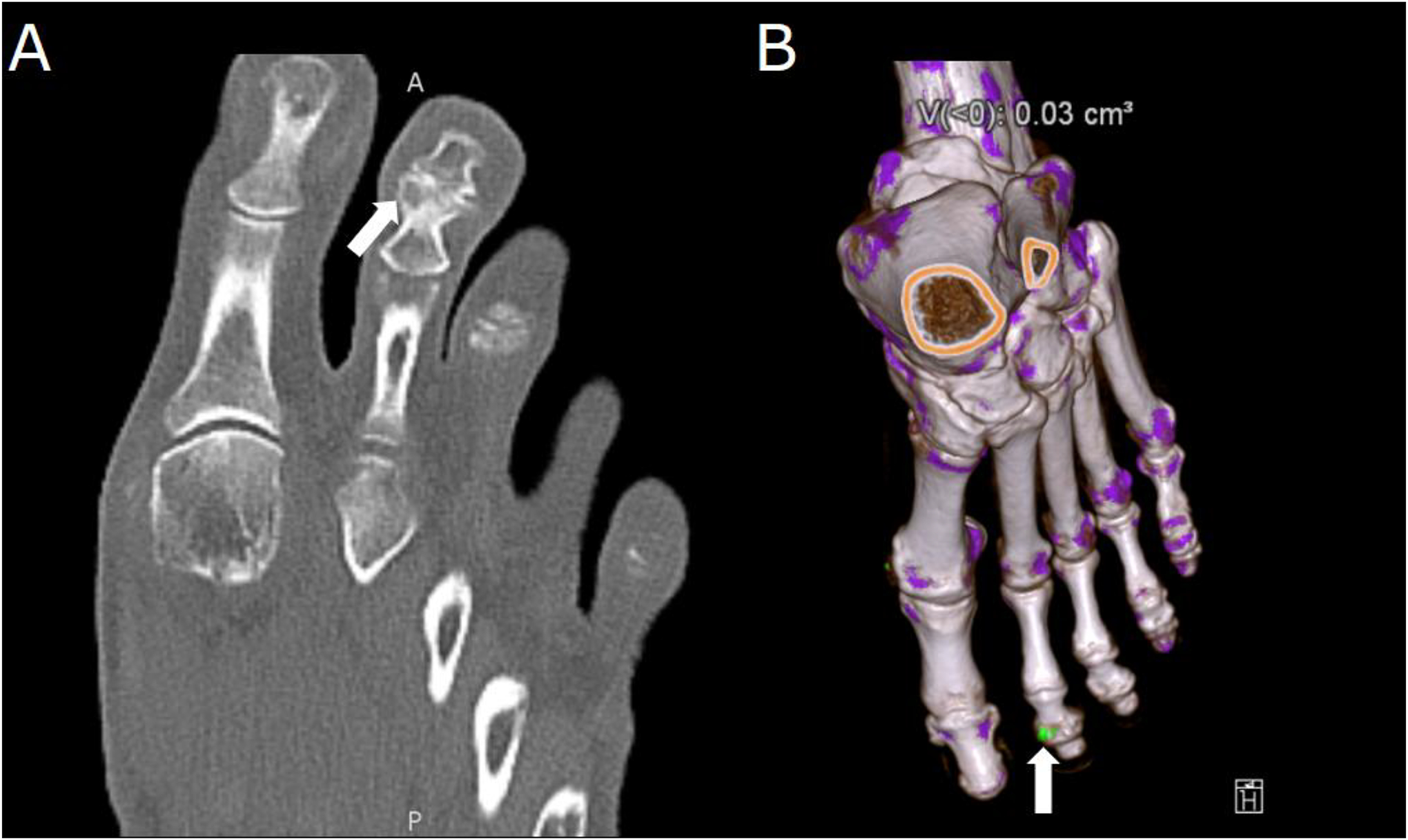
Figure 6.

Dual-energy computed tomography (DECT) of a 65-year-old with polyarticular joint pain and consistently elevated serum uric acid. (A) Demonstration of erosions in the second distal interphalangeal joint characteristic of gout arthropathy (arrow). (B) Material decomposition three-dimensional reconstruction of a DECT acquisition, on which a monosodium urate deposit is seen in green on the same joint (arrow).
