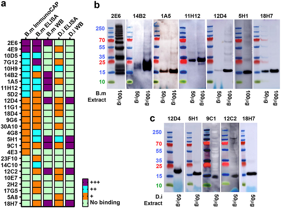
Figure 1. Characterization of IgE mAbs isolated from peripheral blood of subjects with filarial infection.
a, Heatmap representing IgE binding to B. malayi ImmunoCAP (considered positive if >1 kUA/L) and B. malayi and D. immitis binding in ELISA (− no binding detected; + binding 3-10 times background; ++ binding 10-100 times background; +++ binding >100 times background) and immunoblot (WB) (− no binding; +++ presence of a clear band). MAbs are ordered by ImmunoCAP binding value. Data are representative of at least two independent experiments. b, Immunoblot analysis of IgE mAb binding to protein in somatic extracts from B. malayi. c, Analysis as in (b) using D. immitis extracts. For (b) and (c) only immunoblot-positive antibodies are shown.

