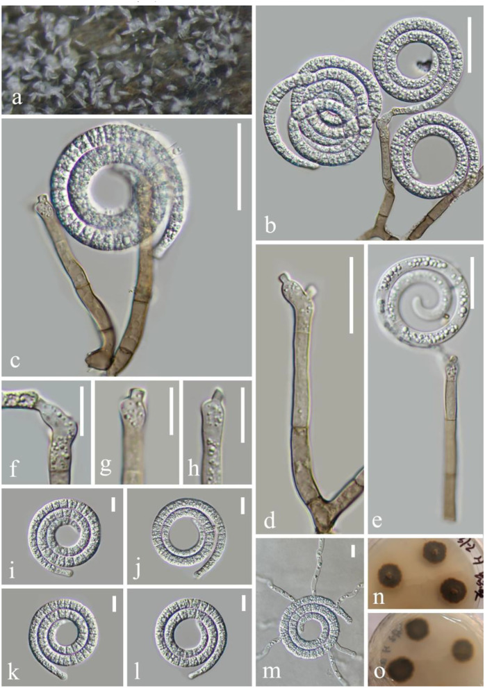
Figure 4.
Neohelicosporium suae (KUN-HKAS 124610, holotype). (a) Colony on decaying wood. (b,c,e) Conidiophores with attached conidia. (d) Conidiophores. (f–h) Conidiogenous cells. (i–l) Conidia. (m) Germinating conidium. (n,o) Colony on PDA observed from above and below. Scale bars: (b,c) 30 μm, (d,e) 20 μm, and (f–m) 10 μm.

