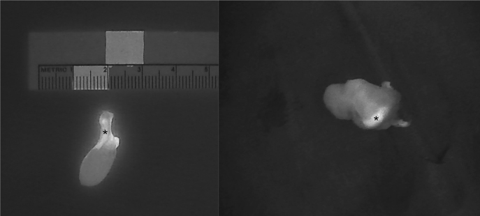
Figure 6:
Intraoperative near-infrared autofluorescence (NIRAF) images of parathyroid adenomas after resection, demonstrating the heterogeneous and less intense fluorescence pattern that differentiates diseased parathyroid glands (PGs) from normal PGs (indicated with *). Frequently, the most intense NIRAF signal comes from residual normal parathyroid tissue in the adenoma.

