
Figure 3. H3.3K27M thalamic gliomas arise from the thalamus proper.
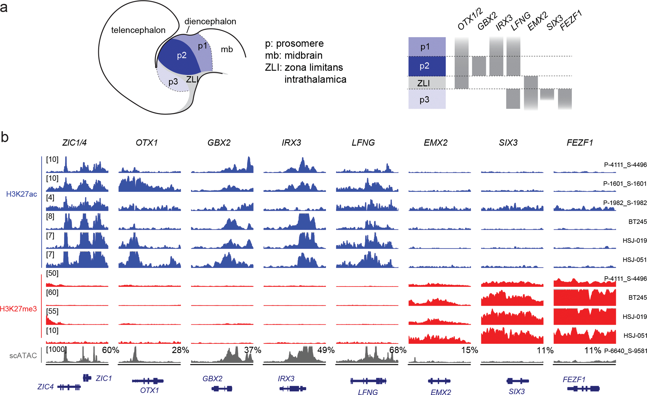
a. Schematic of the developing diencephalon, indicating three embryonal segments (prosomeres, p1–3) and patterning genes that mark each prosomere.
b. H3K27ac and H3K27me3 ChIPseq data and scATACseq for H3.3K27M thalamic HGG primary tumors and cell lines, showing activation of genes marking p2 (the thalamus proper), but silencing of genes marking p3 (the pre-thalamus). Y-axis limit for each sample is indicated in brackets. For scATACseq data, each track represents RPKM-normalized aggregated accessibility for one tumor single-cell population; the percentage of malignant cells where chromatin accessibility in the region was detected (>1 fragment) is indicated for each gene. Chromosome coordinates are indicated in Supplementary Table 15.