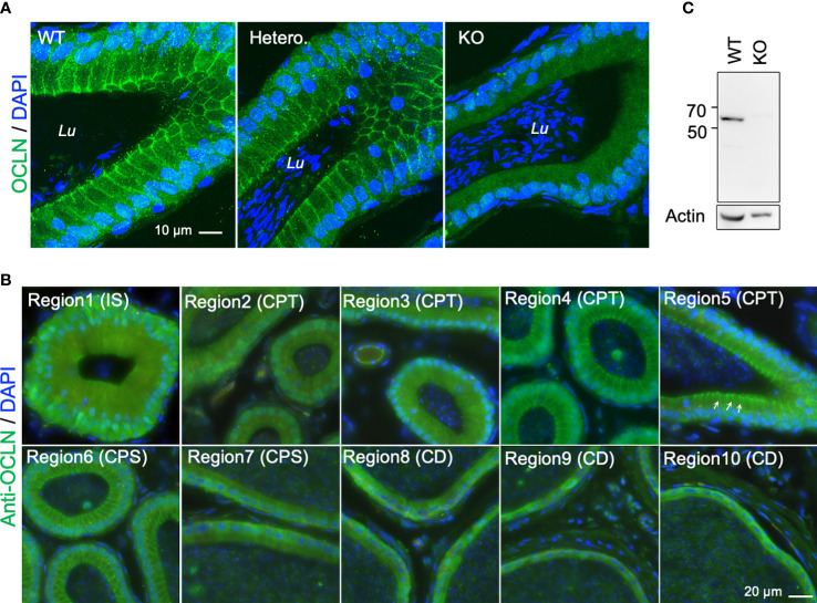
Figure 2.
(A) Typical immunofluorescent-labelling of caput epididymal cryosection immunostaining for anti-OCLN antibody showing enriched OCLN protein in the apical junctional membranes of principal cells in epididymis from WT and heterozygous mice, but absent in Ocln-KO mice. (B) Immunofluorescent-labelling of epididymal cryosection immunostaining for anti-OCLN antibody showing enriched OCLN in region1-6 (IS, CPT and proximal CPS (pCPS) epididymidis). In IS, OCLN is only present in the TJs of principal cells. In the CPT and pCPS principal cells, OCLN also presents in the apical paracellular membranes (arrows). (C) WB-analysis of the same anti-OCLN antibody with KO and WT DC2 cell lysates. Lu, lumen; IS, initial segment; pCPT and dCPT, proximal and distal caput; CPS, corpus; CD, cauda epididymidis; Blue, DNA stained with DAPI.

