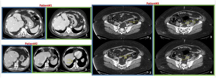
Figure 1.
Radiologic follow-up of patients who experienced disease burden reduction after COVID-19. In blue frames, representative basal CT scans after recovery from COVID-19; in green frames, CT scans at last follow-up or progression. Patient#1. CT scans show absence of liver metastases (section 2 versus section 1). Patient#2. CT scans in green frame show a residual cystic area (section 2) after surgical removal of the lesion at segment VII (section 1) but a new metastatic liver lesions at segment VI (section 3) with the relative measures (16 × 19 mm) (August 2020). Patient#3. Sections 3 and 4 evidence both new and increased size nodules on the peritoneum surface compared to basal sections (1 and 2) (relative measures are reported into the images).
COVID-19, coronavirus disease 19; CT, computed tomography.

