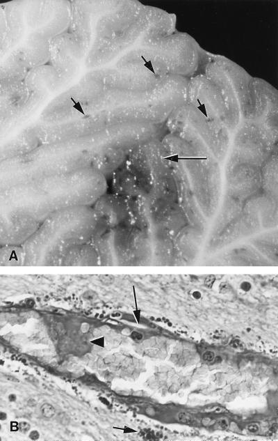
FIG. 1.
Photomicrographs of sections of cerebellum from a suckling piglet necropsied 24 h after inoculation with EHEC strain 86-24. (A) Unstained section showing macroscopic multifocal hemorrhages (short arrows) and necrosis (long arrow) in the medulla and granular layer. (B) PAS section showing endothelial swelling and endothelial necrosis (arrowhead) with subintimal protein insudation (long arrow) in an arteriole and severe perivascular droplet accumulations (short arrow).

