Figure 2.
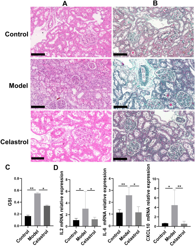
Treatment with celastrol attenuated renal lesions and fibrosis caused by 5/6 nephrectomy. Renal histopathology in each group was subjected to HE and Masson’s trichome staining (A and B). Representative micrographs from each group are shown (BF, bright field, original magnification 200×, Bars = 50 μm). (C) Representative GSI rate for each group. (D) The levels of inflammatory factors IL2 and IL6, as well as chemokine CXCL10 in each group. (A color version of this figure is available in the online journal.)

