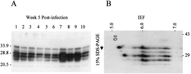
FIG. 4.
Immunoblot analysis of B. turicatae Bdr expression in postinfection clonal populations. Panel A shows the immunoblot of cell lysates of clonal populations (clone designations are indicated above each lane) cultivated for 5 weeks after infection. The lysates were fractionated by sodium dodecyl sulfate-polyacrylamide gel electrophoresis in a 15% gel. In panel B, a cell lysate of a clonal population was fractionated by IEF and two-dimensional sodium dodecyl sulfate-polyacrylamide gel electrophoresis. The pH gradient of the first-dimension IEF tube gel is indicated across the top and was measured with a surface pH probe. One microgram of a tropomysin pI standard (a doublet, 33 and 34 kDa; pI, ∼5.2) was added to each sample and its migration position, as determined by Coomassie staining of the polyvinylidene difluoride blot prior to immunoblot analyses, was circled for reference. Molecular standards are indicated for each panel in kilodaltons. The immunoblots were screened with anti-Bdr antiserum generated in rabbits with recombinant B. afzelii DK1 BdrF1.

