Figure 1.

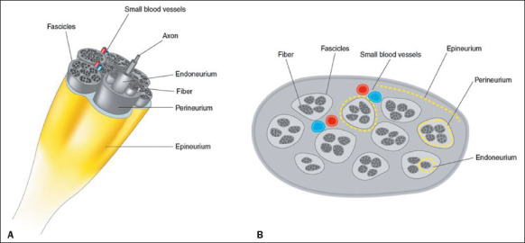
Schematic representations of a normal peripheral nerve and its morphological structures. Three-dimensional view (A) and high-resolution ultrasound cross-sectional view (B), showing the honeycomb pattern.

Schematic representations of a normal peripheral nerve and its morphological structures. Three-dimensional view (A) and high-resolution ultrasound cross-sectional view (B), showing the honeycomb pattern.