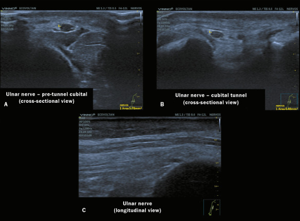
Figure 2.

High-resolution neuromuscular ultrasound images of three sites of the same ulnar nerve obtained with a 4–17 MHz transducer in a VINNO 6 system. A: Image of the ulnar nerve with a normal CSA at the proximal (pre-tunnel) site, showing preserved echogenicity and fascicular pattern. B: Image of a normal ulnar nerve within the cubital tunnel in the right arm. C: Image of the ulnar nerve in a longitudinal view.
