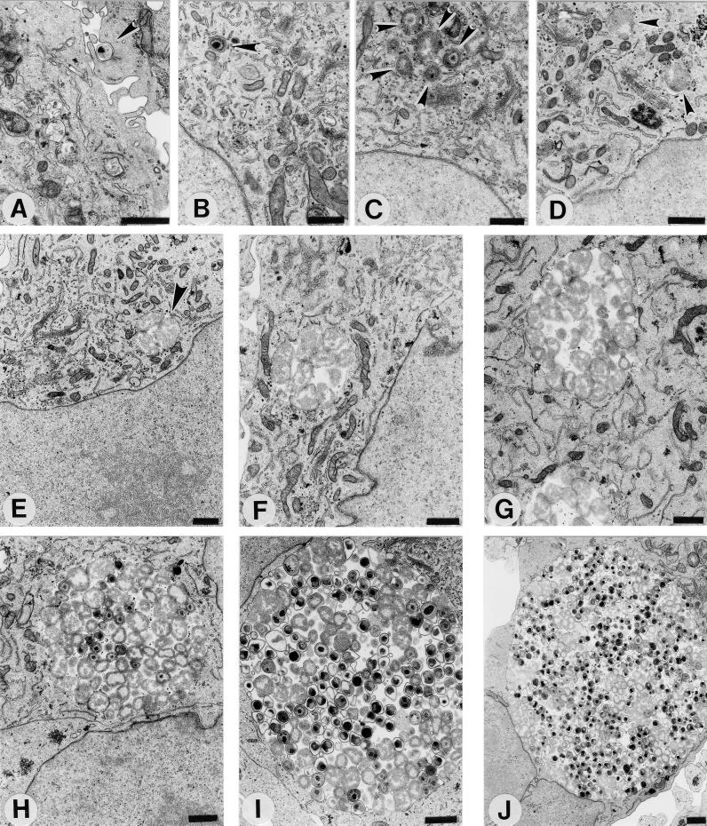
FIG. 1.
The C. pneumoniae AR-39 developmental cycle in HeLa 229 cells. Infected cells were processed for transmission electron microscopy at 0 (A), 2 (B), 8 (C), 12 (D), 19 (E), 24 (F), 36 (G), 48 (H), 60 (I), and 72 (J) h postinfection. Arrowheads indicate intracellular chlamydiae. Scale bars = 1 μm.

