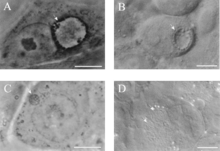
FIG. 5.
Phase- and Nomarski differential-interference-contrast images of C. trachomatis (A and B, respectively) and C. pneumoniae (C and D) inclusions. In comparison to inclusions of C. trachomatis obtained 18 h postinfection, C. pneumoniae inclusions at 36 h postinfection differ in shape and size, although both chlamydiae reach similar stages of their development at these time points postinfection, characterized by the presence of dividing RBs. This difference is most apparent in the presence of clear fluid-filled centers in C. trachomatis inclusions, since C. pneumoniae inclusions are filled with RBs. Scale bars = 10 μm.

