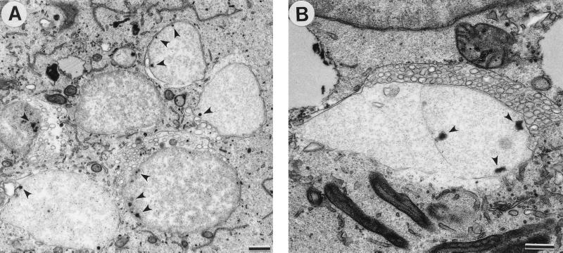
FIG. 6.
Transmission electron micrograph of C. pneumoniae-infected cells treated with ampicillin. (A) Large, abnormal RBs displaying single-cell or no cell division and multiple electron-dense sites, possibly representing nucleoid condensation, near the chlamydial cell wall (arrowheads). (B) Vesicles of unknown function and origin surrounded some giant RBs. Scale bars = 0.5 μm.

