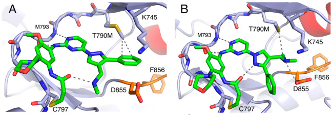
Figure 3.
Lazertinib bound to inactive EGFR(T790M/V948R) with distinct conformations. (A) Lazertinib bound with phenyl ring anchored within van der Waals distance to the T790M methionine (∼3.8 Å) and methane amine involved in intramolecular H-bonding. (B) A “flipped” conformation with outward phenyl and methyleneamine donating an H-bond to D855. Other interactions seen in this structure include an intramolecular dipole–dipole pyrazole to carbonyl (3.2 Å) as well as a van der Waals methyleneamine methyl to thioether (3.8 Å). P-loop cartoon removed for clarity. (PDB ID 7UKW).

