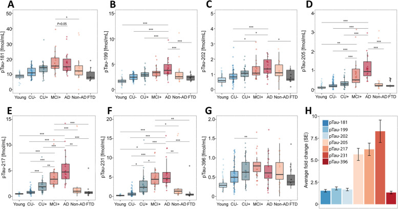
Fig. 1.
Abundances of phosphorylated tau epitopes across the Alzheimer’s disease spectrum in the TRIAD cohort (A-G). The concentrations of the seven phosphorylated tau epitopes, pTau-181 (A), pTau-199 (B), pTau-202 (C), pTau-205 (D), pTau-217 (E), pTau-231 (F) and pTau-396 (G) are plotted for the different groups. The boxplots depict the median (horizontal bar), interquartile range (IQR, hinges) and 1.5 × IQR (whiskers). Group comparisons were computed with a one-way ANCOVA adjusting for age and sex. Tukey honestly significant difference test was used for the post hoc pairwise comparisons in all cohorts. Biomarker fold change between CU- and Alzheimer’s disease groups (H). Abbreviations: Aβ, amyloid-β, AD, Alzheimer’s disease; CU-, Aβ-negative cognitively unimpaired; CU + , Aβ-positive cognitively unimpaired; frontotemporal dementia; MCI + , Aβ-positive mild cognitive impairment; Non-AD, Aβ-negative “AD” dementia or mild cognitive impairment patients. *P < 0.05; **P < 0.01, ***P < 0.001

