
Fig. 1.
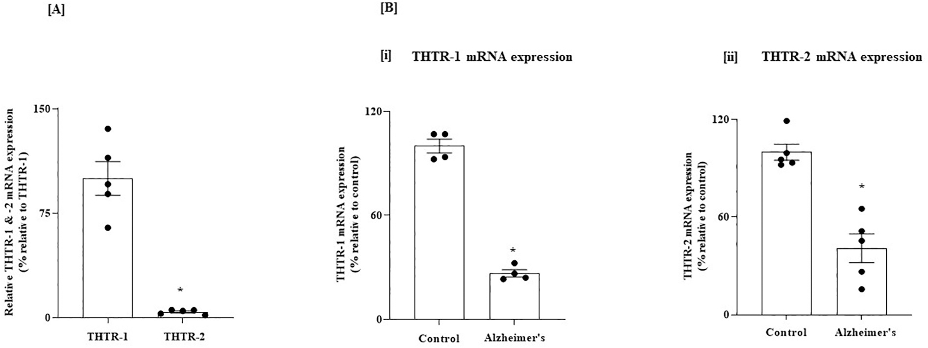
(A) Relative level of expression of THTR-1 and -2 in normal human PFC; and (B) Level of expression of THTR-1 and -2 in PFC of AD and control subjects. (A) PFC tissues from 5 normal subjects were used. (B) PFC tissues from 4 to 5 AD patients and control subjects were used. Level of mRNA expression of THTR-1 (n = 4) and THTR-2 (n = 5) were determined by mean of RT-qPCR; Data were normalized relative to β actin. Statistical analysis was performed using the Student’s t-test. *P < 0.01.