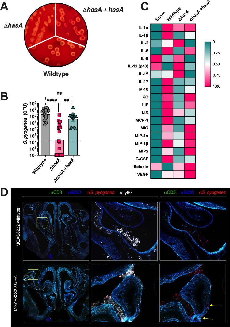
Fig 1. Hyaluronic acid expression by Streptococcus pyogenes promotes nasal infection in B6HLA mice.
(A) S. pyogenes constructs streaked onto TSA + 5% sheep blood agar plates. The plate figure are representative images of the wild-type MGAS8232, ΔhasA mutant and the hasA complemented strain. (B) B6HLA mice were administered ~1×10⁸ CFUs of S. pyogenes MGAS8232 wildtype, ΔhasA, or ΔhasA +hasA strains intranasally and sacrificed 48 h later. Data points represent CFUs from cNTs of individual B6HLA mice. Horizontal bars represent the geometric mean. Significance was determined by Kruskal Wallis one-way ANOVA with Dunn’s multiple comparisons test (****, P < 0.0001; **, P < 0.01). The horizontal dotted line indicates the theoretical limit of detection. (C) Heat-map of cytokine responses in cNTs of B6HLA mice during S. pyogenes infection. Data shown represent normalized median cytokine responses from cNTs (n ≥ 3 per group). (D) Immunohistochemistry of infected cNTs at 24 h post-infection with wildtype S. pyogenes MGAS8232 and the ΔhasA mutant. Sections were stained with α-S. pyogenes (red), αB220 (blue), αCD3 (green), and αLy6G (white) antibodies. Panels are a close-up view from the boxed section. Arrows indicate regions with internalized S. pyogenes.

