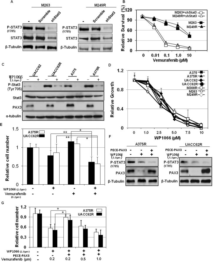
Figure 6. Inhibition of Stat3 overcomes the acquired resistance to vemurafenib in resistant melanoma cells.
(A) M263R and M249R melanoma cells stably expressing control shRNA (shScr), shStat3 or shPAX3 were stimulated different doses of vemurafenib as indicated. (C) Sensitive and resistant A375 and UACC62 melanoma cells were stimulated with 5.0μm WP1066. Total protein was collected at 6 hr after stimulation. Protein expression of Stat3 and phospho-Stat3 were analyzed by western blot along with tubulin, which served as a loading control. A375R and UACC62R melanoma cells were stimulated with different doses of vemurafenib (B), WP1066 (D) or both (E) as indicated. Melanoma cell growth was assessed by MTT assays. Relative growth (RG) was calculated as the ratio of treated to untreated cells at each dose for each replicate (*p< 0.01; **p<0.05). A375 and UACC62 resistant cells with PECE-PAX3 plasmids were stimulated with vemurafenib and/or WP1066 as indicated. (F) Total protein was collected 6 hr after stimulation. Protein expression of phospho-Stat3 and PAX3 were analyzed by western blot along with tubulin, which served as a loading control. (G) Cells were subjected to MTT assays to evaluate the relative cell numbers. Relative growth (RG) was calculated as the ratio of treated to untreated cells at each dose for each replicate (*p< 0.01).

