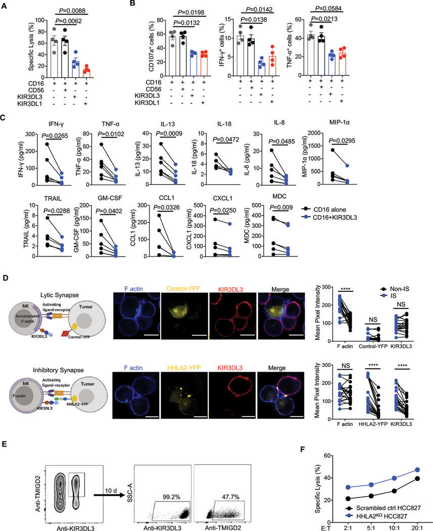
Fig. 4. KIR3DL3 inhibited primary NK function and mediates HHLA2+ tumor immune resistance.
(A-C) Primary KIR3DL3+ NK cells redirected cytotoxicity assay against P815. (A) The lysis of P815 cells (n=4). (B) The degranulation (CD107a) and cytokine production (IFN-γ and TNF-α) of primary KIR3DL3+ NK cells (n=4). CD56 and KIR3DL1 served as a negative and positive control, respectively. (C) Cytokine production in the co-culture supernatant of primary KIR3DL3+ NK cells with indicated antibody-coated P815 (n=4–6). Data are represented as mean ± SD.
(D) HHLA2-KIR3DL3 mediated inhibitory synapses formation. Left: The cartoons depicting lytic and inhibitory synapses. Middle: Representative confocal images of cell conjugates acquired after KIR3DL3+ NK92 cell-A427 contact followed by staining with anti-KIR3DL3 and Phalloidin. Scale bars, 10 𝜇m. Right: The intensity quantification of F-actin, YFP, and KIR3DL3 at the immunological synapses (IS) and the cell surface away from synapses (Non-IS) from NK92 cell-Control/A427 (n=20) and NK92-HHLA2/A427 (n=21) conjugates. Mouse Tim-3-YFP/A427 was used as the control.
(E) Primary KIR3DL3+ TMIGD2+ NK cells were sorted and cultured in vitro. Ten days later, KIR3DL3 and TMIGD2 expression was examined by flow cytometry.
(F) Lysis of scrambled control or HHLA2KO HCC827 by primary KIR3DL3+ TMIGD2+ NK cells in (E) at indicated E:T ratios. Data are mean for duplicate measurements. Data are representative of three independent experiments with three different donors.
P values by a one-way ANOVA (A-B) or two-tailed paired (C-D). ****P < 0.0001; NS, not significant.

