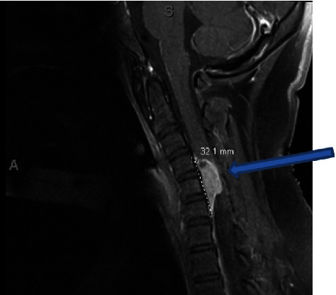
Figure 1.

Sagittal T1-weighted preoperative MRI with contrast showing an anterior intradural extramedullary mass, measuring 1.6 × 0.9 × 2.8 cm (blue arrow). The mass is centered at C5 and spans from C4-6 with what appears to be a dural tail. The mass shows significant compression of the spinal cord.
