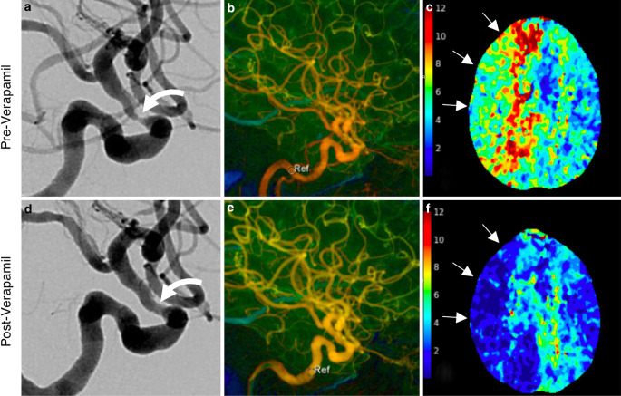
Fig. 3.
Calcium channel blocker infusion as monotherapy for intracranial giant cell arteritis. Pre-treatment angiography (lateral RICA projection) shows severe focal supraclinoid segment stenosis (a). Color-coded four-dimensional DSA (4D-DSA, b) shows prolonged transit time throughout the RICA circulation; sample velocity at the petrous segment time-to-peak (TTP) velocity of 4.53s. CTP performed the day prior to intervention showed at-risk tissue (prolonged Tmax) throughout the right ICA territory (c, arrows). Post-verapamil infusion (20 mg, 15 min delay) angiogram is shown in d, with significant improvement in lumen diameter. Post-verapamil 4D-DSA (e) shows improved flow throughout the ICA distribution and normalization of TTP in the petrous segment (1.0s) (circle, labeled Ref). A CTP performed 10 h after verapamil infusion shows durable improvement in Tmax in RICA distribution (f)

