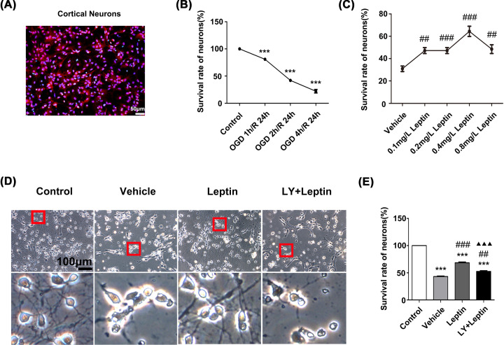
Figure 1. Leptin protected primary cortical neurons against OGD/R injury.
(A) Cortical neurons were labeled with NeuN and DAPI. (B) The viability of neurons that were subjected to OGD for different degrees (0, 1, 2, 4 h) followed by reoxygenation for 24 h. (C) The viability of neurons that were treated with different concentrations of leptin (0, 0.1, 0.2, 0.4 or 0.8 mg/L) and subjected to OGD for 2 h/R for 24 h. (D) Representative images of neurons in each group under a microscope (scale bar: 100 μm). (E) The viability of cortical neurons in each group. The data are presented as the means ± SEMs (n=3). ***P<0.001, versus the control group; ##P<0.01, versus the vehicle group; ###P<0.001, versus the vehicle group; ▲▲▲P<0.001, versus the leptin group.

