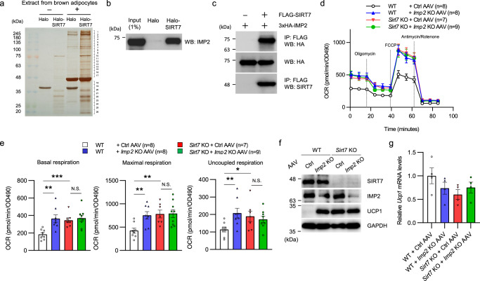
Fig. 7. SIRT7 suppresses energy expenditure via IMP2 in brown adipocytes.
a Halo-SIRT7 pull-down assay with extracts from brown adipocytes. Eluted proteins were resolved by SDS-PAGE, followed by silver staining. The gel lanes were cut into 26 pieces and the proteins within each gel piece were analyzed by mass spectrometry. b Halo-SIRT7 pull-down assay performed with lysates from HEK293T cells overexpressing 3×HA-IMP2. Overexpressed IMP2 was detected by WB with an anti-IMP2 antibody. c Co-IP assay detecting the interaction between FLAG-SIRT7 and 3×HA-IMP2 in HEK293T cells. d, e The effect of Imp2 deficiency on mitochondrial respiration in differentiated primary brown adipocytes from WT and Sirt7 KO mice. SVF cells were infected with the indicated recombinant AAV and differentiated for 9 days. Time course OCR (d) and quantification of mitochondrial respiration (e). p = 0.0020 (WT + Ctrl AAV vs. WT + Imp2 KO AAV), p = 0.0005 (WT + Ctrl AAV vs. Sirt7 KO + Ctrl AAV) in basal respiration; p = 0.0039 (WT + Ctrl AAV vs. WT + Imp2 KO AAV), p = 0.0031 (WT + Ctrl AAV vs. Sirt7 KO + Ctrl AAV) in maximal respiration; p = 0.0099 (WT + Ctrl AAV vs. WT + Imp2 KO AAV), p = 0.0348 (WT + Ctrl AAV vs. Sirt7 KO + Ctrl AAV) in uncoupled respiration. f, g Western blot (f) and real-time qPCR (g) analysis of Ucp1 in the cells described in (d). n = 4 independent samples per group in (g). WB western blotting, IP immunoprecipitation, N.S. not significant. Data are presented as means ± SEM. All numbers (n) are biologically independent samples. The screening experiment (a) were performed one time. *p < 0.05, **p < 0.01 by two-tailed Student’s t-test. Source data are provided as a Source Data file.

