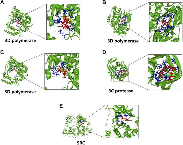
FIGURE 8.
Molecular docking models of compounds with potential protein targets. (A–C) Hexadecamethyl cyclooctasiloxane, 3-thiazolidinecarboxylic acid and monobutyl phthalate bind to EV71 3D polymerase, respectively. (D) Diheptyl phthalate binds to EV71 3C protease. (E) Nona-2,3-dienoic acid, ethyl ester binds to SRC. The left side of all the panels show the whole models, the right side shows the zoomed-in display of the binding site. The proteins are shown in main chain ribbon mode (green), the compounds are shown in ball and sticks mode (red), and the amino acids residues participate in binding are shown in stick mode (blue) with labels.

