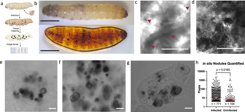
Fig. 3. Using tissue clearing to visualize the melanin-based immune response against C. neoformans in situ.
Using tissue-clearing techniques (a), we allowed for better visualization of melanized nodules within the intact G. mellonella, as seen in a before (top) and after (bottom) view of the same infected larvae (b). Under microscopy, the uninfected larvae had little to no melanized nodules (red arrows indicate dark, irregular, or opaque areas in the tissue) (c), whereas the C. neoformans larvae had very clear and distinct nodules (d). These nodules were viewed under high magnification, where the nodule structure and encapsulated fungus were apparent (e, f). These structures were very similar in appearance to the nodules extracted from hemolymph (g). h The size of the melanized nodules were quantified using particle measuring software. Infected particles represent n = 771 independent particles, uninfected particles represent n = 104 independent particles, (Mann–Whitney test, p = 0.016). Error bars signify median with 95% Confidence Interval. i The melanized nodules of C. neoformans appeared to have no tissue tropism, but were found in distinct clumps or areas throughout the larvae (red arrows). b–g, i are representative images from three biological replicates. Scale bars in e–g, i represent 10 µm, c, d represent 500 µm, and b represent 50 mm.

