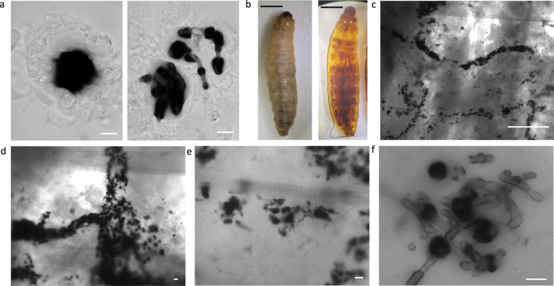
Fig. 4. Visualizing the melanization response to infection with Candida albicans.
a Similar to the hemolymph from larvae infected with C. neoformans, we are able to see melanized nodules within the hemolymph. The melanized spot within these nodules appears more diffuse/less defined, and in some, the presence of less-melanized hyphae is distinct (red arrows). b We can also use tissue clearing to visualize the melanized nodules during C. albicans infection. c, d The melanized C. albicans seem to cluster in specific areas, in long strips within the larvae (yellow arrows). e, f Under higher magnification, we can see hyphal structures of C. albicans, which corresponds to what is previously known about C. albicans morphology in G. mellonella. Interestingly, the hyphae appear less melanized (red arrows) compared to the spherical yeast (white arrows) (f). All panels show representative images from 3 biological replicates. Scale bars in a, d–f represent 10 µm, in c represent 500 µm, and in b represent 50 mm.

