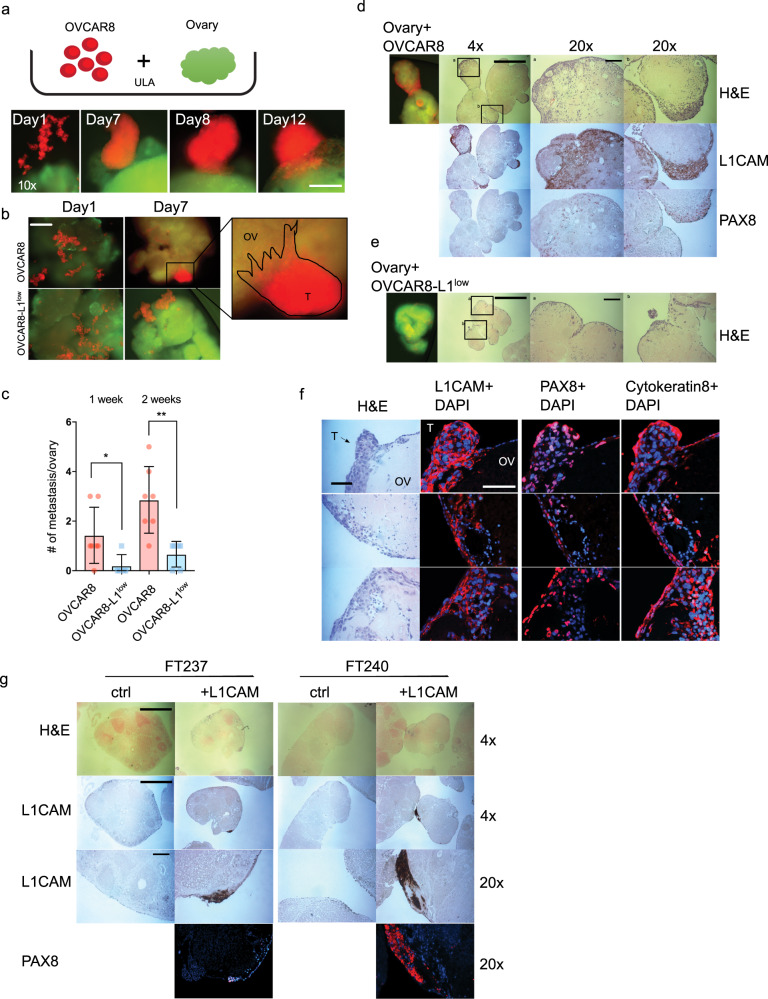
Fig. 8. L1CAM is important for ovary invasion.
a Upper row: Experiment schematic of co-culture of cells with an isolated mouse ovary under ULA culturing conditions. Lower row: representative observed invasion sequence over time. Scalebar represents 100 µm. b Representative fluorescent image after one week’s co-culture of OVCAR8 or OVCAR8-L1low (red) with isolated ovary (green) under ULA culture conditions. Scalebar represents 500 µm. c Bar graph representation of various OVCAR8 cells metastases to the ovary (more than 10 cells) after 1 and 2 weeks of co-culture. Three biological experiments were performed with at least 3 ovaries for analysis. d, e Fluorescent and IHC images representing co-culture of OVCAR8 or OVCAR8-L1low with ovary, respectively. f Fluorescent images of ovaries co-cultured with OVCAR8wt for expression of L1CAM, PAX8 and Cytokeratin 8. Scalebar represents 100 µm. g H&E and IHC images representing ovaries co-cultured with FT237 or FT246 transfected with L1CAM or a control vector. d, e and g Scalebar represents 100 µm for 20x objective and 1000 µm for 4x objective.

