
FIGURE 8.
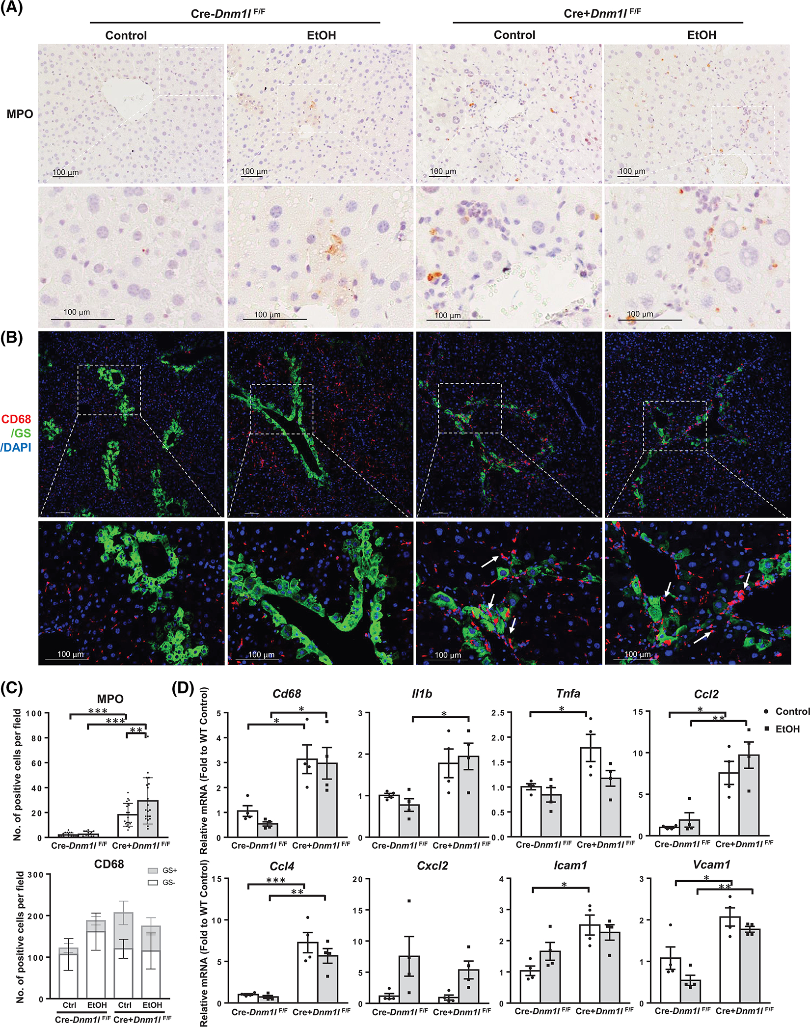
Loss of dynamin-related protein 1 (DRP1) promotes immune cell infiltration in mouse liver. (A) Representative images of immunohistochemistry staining for liver MPO of indicated mice. (B) Representative images of immunofluorescence staining of liver CD68 (red) and glutamine synthetase (GS, green) of indicated mice. Arrows denote macrophages clustered in the central vein region. (C) Quantification of (A) and (B). Data are presented as means ± SD (n = 15 fields from 3 mice). (D) Quantitative real-time polymerase chain reaction analysis of inflammation genes in mouse liver. Data are presented as means ± SEM (n = 4). *p < 0.05, **p < 0.01, ***p < 0.001; one-way analysis of variance analysis with Bonferroni’s post hoc test. DAPI, 4’,6-diamidino-2-phenylindole; EtOH, ethanol; MPO, myeloperoxidase.