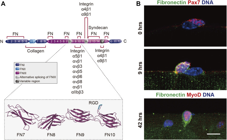
FIGURE 5.
Autologous expression of fibronectin by MuSCs. (A) Scheme showing the domain structure of fibronectin and the binding sites for collagen, integrin and syndecan. The insert shows the seventh through the RGD-containing 10th type III repeats of fibronectin (Leahy et al., 1996) obtained from the RCSB Protein Data Bank (Berman et al., 2000). The RGD motif is highlighted in blue. (B) Immunostaining showing the endogenous expression of fibronectin (green), Pax7 or MyoD (red) and DNA (blue) in muscle stem cells on enzymatically isolated mouse single muscle fibers after 0, 9, and 42 h (hrs) in culture. Scale bar = 20 µm.

