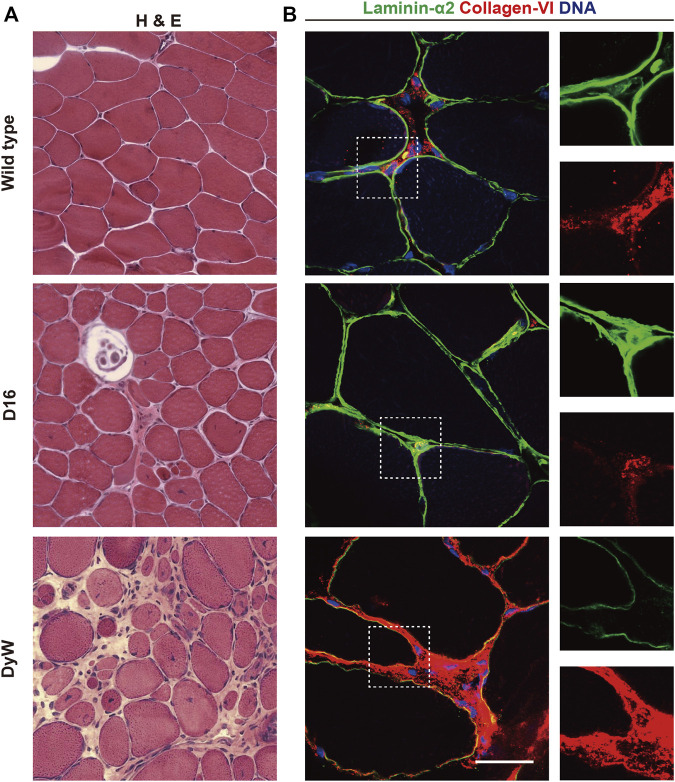
FIGURE 7.
ECM and muscular dystrophy. (A) Hematoxylin and eosin (H&E) staining of skeletal muscle sections from wild type mice, COL6A3 mutant D16 mice, and LAMA2 mutant dyW mice (Kuang et al., 1998; Pan et al., 2014). (B) Immunostaining of skeletal muscle cross sections from wild type, D16 and dyW mice showing laminin α2 (green), collagen VI (red) and DNA (blue).

