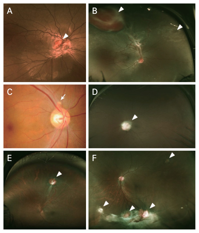
Fig. 1.
A variety of clinical features of retinal capillary hemangioblastoma (RCH) in this study. (A) Fundus photograph of a 20-year-old male patient with von Hippel-Lindau (VHL) disease at the initial visit. Juxtapapillary RCH (arrowhead) with macular epiretinal membrane and subretinal fluid were observed. He had the p.Trp88Ter mutation of VHL gene. (B) Fundus photograph of a 23-year-old female patient with VHL disease at the initial visit. Arrowheads indicate RCH. Large peripheral RCH (left arrowhead) with tortuous feeding artery, dilated draining vein, and glial proliferation were observed. (C) Fundus photograph of a 46-year-old male patient with VHL disease at a year after diagnosis. A small juxtapapillary RCH (arrow) was observed. He had the p.Glu70Lys mutation of VHL gene. (D) Fundus photograph of a 42-year-old male patient with VHL disease at the initial visit. Juxtapapillary RCH (arrowhead) covering the optic disc was observed. (E) Fundus photograph of a 24-year-old female patient without VHL disease at the initial visit. Peripheral RCH (arrowhead) with tortuous feeding artery, dilated draining vein, and prominent glial proliferation were observed. (F) Fundus photograph of a 42-year-old male patient with VHL disease at 12 years after diagnosis. Peripheral RCHs (arrowheads) with inferior tractional retinal detachment were observed. Right after this examination, he underwent four vitrectomy surgery in 4 years. On his last visit, his retina of the left eye was flat and best-corrected visual acuity of the left eye was 0.4 logarithm of the minimum angle of resolution.

