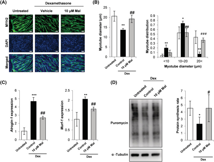
Figure 1.

(A) Myosin heavy chain (MYH2) immunocytochemistry of C2C12 myoblasts cultured as follows: (1) differentiation media (DM) for 120 h (untreated); (2) DM for 96 h and DM plus 10 μM Dex for 24 h; (3) DM for 96 h and DM plus 10 μM Dex and 10 μM malotilate (Mal) for 24 h (scale bar = 100 μm). (B) Myotube diameter and myotube diameter distribution. (C) qPCR analysis of atrogin‐1 and MuRF‐1 expression. (D) SUnSET assay of protein synthesis. α‐Tubulin expression was used as the loading control. *P < 0.05, **P< 0.01, ***P < 0.001 compared to untreated. # P < 0.05, ## P < 0.01, ### P < 0.001 compared with Dex treated.
