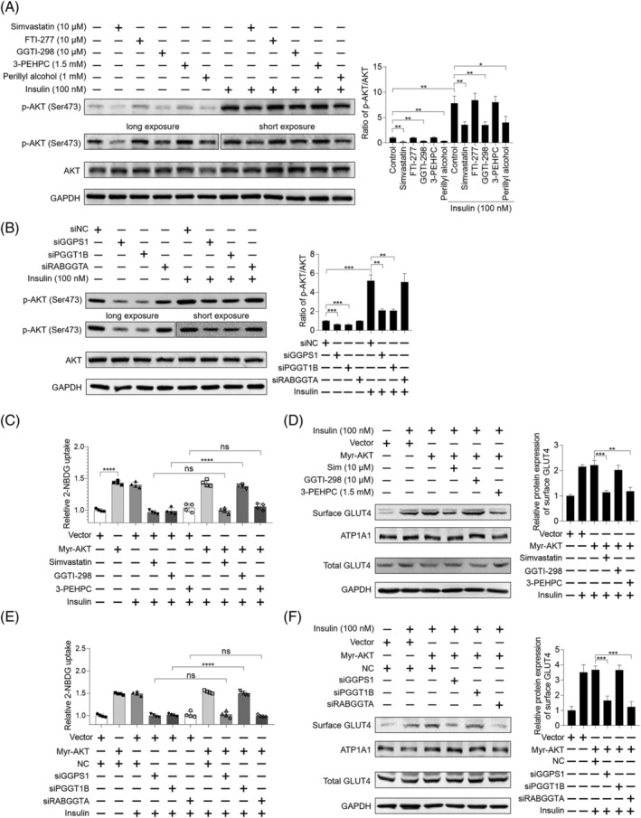
Figure 3.

Insulin signalling is not necessary for simvastatin‐caused inhibition of insulin‐stimulated glucose uptake in skeletal muscle cells. (A) C2C12 myotubes were pretreated with 10 μM simvastatin, 10 μM FTI‐277, 10 μM GGTI‐298, 1.5 mM 3‐PEHPC, and 1 mM perillyl alcohol for 24 h. Then cells were incubated with or without 100 nM insulin for 30 min, total protein was harvested, and the expression of indicated proteins was analysed by western blot, with GAPDH as the loading control (n = 3). Long exposure of p‐AKT (Ser473) for 30 s and short exposure of p‐AKT (Ser473) for 5 s were shown. (B) C2C12 myotubes were previously transfected with siRNAs targeting GGPS1, PGGT1B, and RABGGTA, respectively, for 48 h. Then cells were incubated with or without 100 nM insulin for 30 min, total protein was harvested, and the expression of indicated proteins was analysed by western blot, with GAPDH as the loading control (n = 3). Long exposure of p‐AKT (Ser473) for 30 s and short exposure of p‐AKT (Ser473) for 5 s were shown. (C) C2C12 myotubes were previously transfected with or without 1 μg Myr‐AKT plasmid using Lipofectamine 3000 for 48 h. Then the cells were treated with 10 μM simvastatin, 10 μM FTI‐277, 10 μM GGTI‐298, 1.5 mM 3‐PEHPC, and 1 mM perillyl alcohol for another 24 h. Cells were exposed to 2‐NBDG containing 100 nM insulin for 30 min, and 2‐NBDG uptake was measured by fluorescence detection (n = 3). (D) C2C12 myotubes were previously transfected with 1 μg Myr‐AKT plasmid using Lipofectamine 3000 for 48 h. Then the cells were treated with 10 μM simvastatin, 10 μM FTI‐277, 10 μM GGTI‐298, 1.5 mM 3‐PEHPC, and 1 mM perillyl alcohol for another 24 h. Cells were incubated with 100 nM insulin for 30 min, then protein samples were harvested, and the expression of indicated proteins was checked by western blot, with GAPDH as the loading control (n = 3). (E) C2C12 myotubes were previously transfected with or without 1 μg Myr‐AKT plasmid using Lipofectamine 3000 for 48 h. Then cells were transfected with siRNAs targeting GGPS1, PGGT1B, and RABGGTA, respectively, for another 48 h. Cells were exposed to 2‐NBDG containing 100 nM insulin for 30 min, and 2‐NBDG uptake was measured by fluorescence detection (n = 3). (F) C2C12 myotubes were previously transfected with or without 1 μg Myr‐AKT plasmid using Lipofectamine 3000 for 48 h. Then cells were transfected with siRNAs targeting GGPS1, PGGT1B, and RABGGTA, respectively, for another 48 h. Cells were incubated with 100 nM insulin for 30 min, protein samples were harvested, and the expression of indicated proteins was checked by western blot, with GAPDH as the loading control (n = 3). Data represented the mean ± SEM. Statistical analysis was performed with one‐way ANOVA. *P < 0.05; **P < 0.01; ***P < 0.001; ****P < 0.0001; ns denotes no significance.
