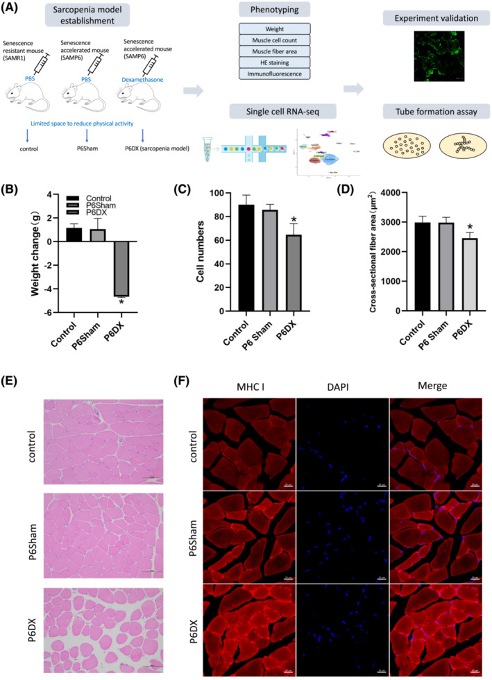
Figure 1.

Establishment and evaluation of a murine sarcopenia model. (A) A scheme to illustrate the workflow of this study. A mouse model for sarcopenia is established and evaluated by different criteria. TA biopsies from sarcopenia mice (n = 5) as well as from mice in two other control groups (n = 5 each group) are subject to single‐cell transcriptome analysis. Findings from scRNA‐seq are validated by in vivo and in vitro experiments. (B–D) Criteria to evaluate sarcopenia mice model. The body weights, muscle cell counts and cross‐sectional fibre area are significantly decreased in P6DX sarcopenia mice group during the sarcopenia establishment procedure. (E) HE‐stained TA muscle of R1 control, P6Sham and P6DX groups; the muscle cell structures are loose in P6DX sarcopenia sample. Scale bar represents 100 μm. (F) Immunofluorescence staining of the three experimental groups. Myosin heavy chain I (MCH I) is increased in P6DX sarcopenia sample. Scale bar represents 20 μm.
