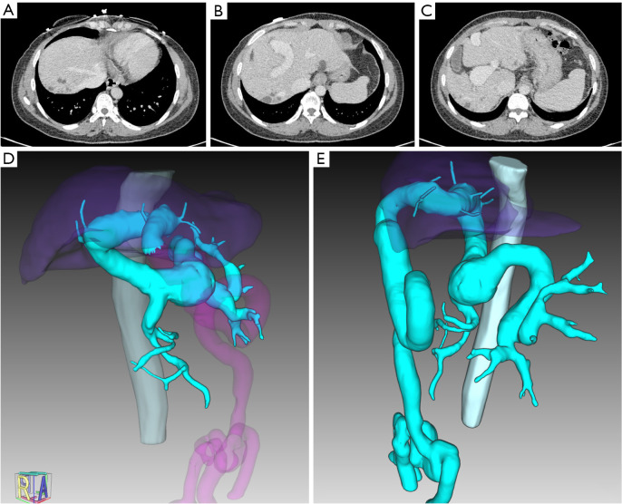
Figure 1.
Imaging of abnormal intrahepatic portal vein without second-order branches. (A-C) Contrast-enhanced CT scans on portal venous phase showed a dilated portal trunk and absence of bifurcation. (D,E) Images of 3D reconstruction of portal vein and branches (vein in cyan). CT, computed tomography.

