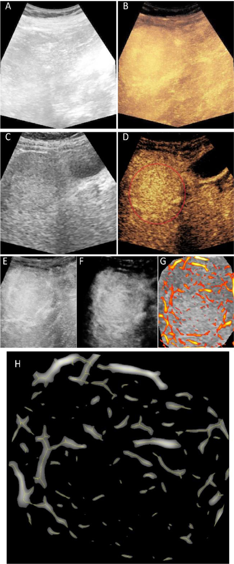
Fig. 1.

Representative outcome from image processing steps. The maximum intensity projection (MIP) images from original B-mode ultrasound (US) (A) and contrast-enhanced US (CEUS) (B) image sequences. Region-of-interest (ROI) selection from a single B-mode US (C) and CEUS (D) image. Motion corrected version of the CEUS-MIP (E) and the result of clutter filtering (F). Enhanced tubular structures overlaid on original and resized B-mode US reference for only the tumor region (G). Centerlines of the vessel on enlarged tumor area used for morphological feature extraction (H).
