Fig. 1.
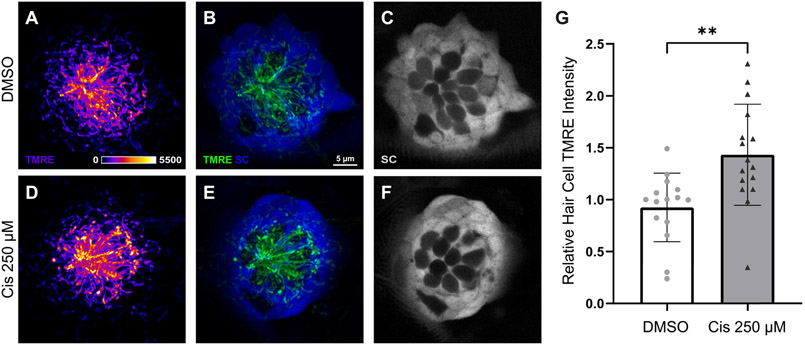
Hair cell mitochondrial activity increases following cisplatin exposure. Maximum-intensity projections of confocal images show TMRE-loaded Tg(TNKS1bp1:EGFP) zebrafish neuromasts with GFP-expressing supporting cells (SC) and GFP-negative hair cells after 2 h treatment with 0.1% DMSO (A-C) and 250 μM cisplatin (D – F). TMRE labeling is observed most prominently in hair cells (A-B; d-E). Mean hair cell TMRE intensity (normalized to control) was elevated in the cisplatin-exposed neuromasts (G, **p = 0.0021, unpaired t-test). n = 15 – 16 zebrafish. N = 4 experimental trials. Error bars = standard deviation.

