Fig. 2.
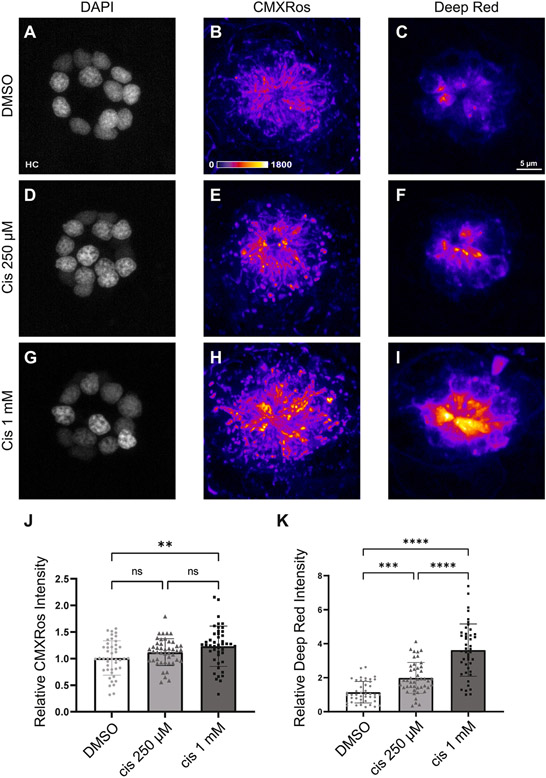
The degree of cisplatin-induced mitochondrial hyperpolarization is dose dependent. Maximum-intensity projections of confocal images show neuromasts treated with 0.1% DMSO (A – C), 250 μM cisplatin (D – F), and 1 mM cisplatin (G – I). Hair cells were labeled with DAPI and mitochondria were stained with Mitotracker CMXRos and MitoTracker Deep Red. Mean intensities (normalized to control) increased in a dose-dependent manner across both MitoTracker CMXRos (J, p = 0.0058, one-way ANOVA) and MitoTracker Deep Red (K, p < 0.0001, one-way ANOVA) indicators. Post-hoc analysis with Tukey’s multiple comparisons test failed to detect significant changes in mean CMXRos intensities between DMSO and 250 μM cisplatin (J, ns = 0.23) and 250 μM cisplatin and 1 mM cisplatin (J, ns = 0.24). For MitoTracker Deep Red, significant differences were observed using Tukey’s multiple comparisons test between all treatment groups (K, **p = 0.0011, p < 0.0001). n = 135 neuromasts. N = 3 experimental trials. Error bars = standard deviation.

