Fig. 6.

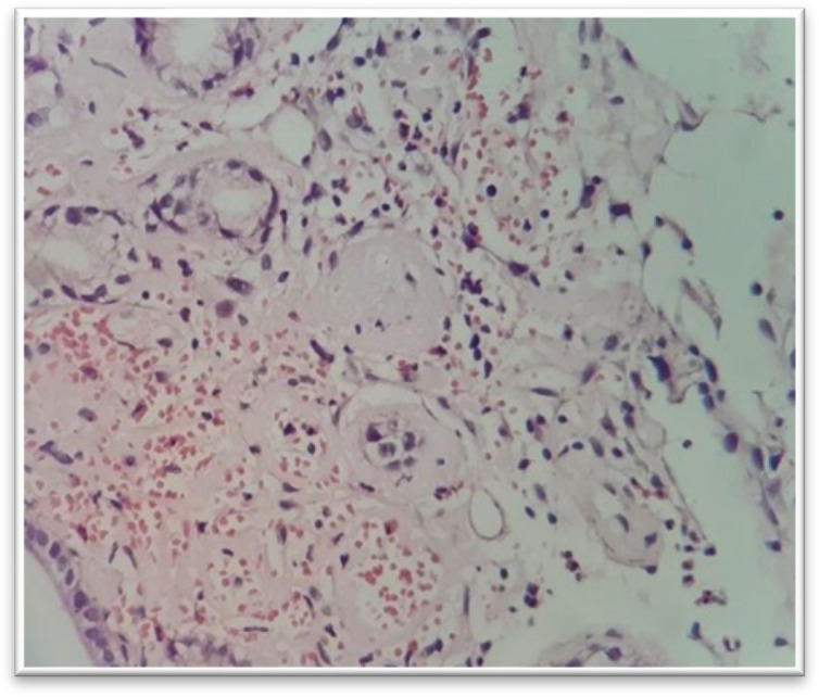
Amyloidosis, H&E stain, biopsy shows amorphous eosinophilic material present around small blood vessels of the mucosa. In the Congo red stain, the material demonstrates an apple-green birefringence (40 x)

Amyloidosis, H&E stain, biopsy shows amorphous eosinophilic material present around small blood vessels of the mucosa. In the Congo red stain, the material demonstrates an apple-green birefringence (40 x)