FIGURE 2.
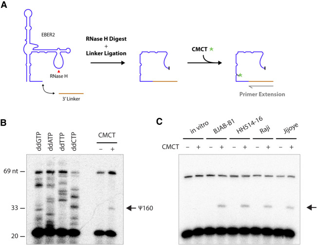
Detection of EBER2 pseudouridylation by primer extension. (A) Experimental outline for verifying pseudouridylation by primer extension assay following CMCT-treatment. A linker is ligated to the 3′ end of EBER2, which is then digested with RNase H at the major loop region (red arrowhead). EBER2 is treated with CMCT to form a covalent adduct at pseudouridylation sites that will elicit a premature stop in reverse-transcription reactions. (B) A representative primer extension assay with digested EBER2 is shown, which corroborates the presence of pseudouridylation (arrow). Sequencing ladders utilizing dideoxy nucleotides are included for orientation. Please note that not each nucleotide of EBER2 is displayed at equal band intensity, which is a common observation with direct RNA sequencing using reverse transcription and dideoxy nucleotides. (C) EBER2 pseudouridylation is found in a wide array of EBV-positive cell lines. EBER2 isolated from BJAB-B1, HH514-16, Raji, and Jijoye cells was subjected to CMCT treatment followed by primer extension assay. In vitro-transcribed EBER2 was also examined as a negative control.

