
Figure 2. Dopamine transporter uptake in basal ganglia shown by SPECT or PET.
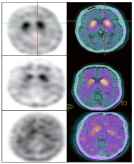
Dopamine transporter uptake in basal ganglia demonstrated by TRODAT-1 SPECT imaging. The superior row shows a normal uptake in the caudate and putamen. The medium row shows an asymmetric uptake in a PD patient. The lower row shows a bilateral minimal uptake in a DLB patient. (Reproduced with permission from Dr. Artur Coutinho, Nuclear Medicine Center - Inrad - HC-FMUSP).