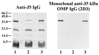
FIG. 1.
Immunoblot of OmpA-deficient bacteria. Bacteria were grown to mid-log phase and then boiled in SDS-PAGE sample buffer. Bacterial lysates were then electrophoresed on SDS–16% polyacrylamide gels and transferred to nitrocellulose. Staining antibodies included polyclonal rabbit anti-J5 IgG (left) and a monoclonal antibody directed to the 35-kDa OMP (2D3; right). Bacterial strains: wild-type OmpA+ E. coli O18:K1:H7 (lane 1); E91, an OmpA-deleted mutant of E. coli O18:K1:H7 (lane 2); E69, an OmpA-restored mutant of E. coli O18:K1:H7 (lane 3). Positions of molecular weight markers (in kilodaltons) are shown at the left.

