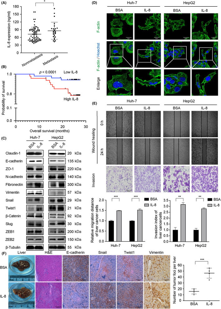
FIGURE 1.

Interleukin‐8 (IL‐8) promotes metastasis of liver cancer and predicts patient outcomes. (A) Comparison of serum interleukin‐8 (IL‐8) expression levels between hepatocellular carcinoma (HCC) metastatic and nonmetastatic groups (p = 0.05). (B) HCC patients with low IL‐8 expression had better OS. (C) Expression of Snail, Twist1, N‐cadherin, fibronectin, and vimentin increased, whereas E‐cadherin and ZO‐1 decreased in liver cancer cells treated with rh‐IL‐8. (D) A spike‐like filopodia morphology change was observed in IL‐8‐stimulated cells. Cells were stained with F‐actin (green) and counterstained with Hoechst (blue). Magnification, ×400. (E) Wound healing and Transwell assays in Huh‐7 and HepG2 cells. IL‐8 stimulation enhanced liver cancer cell migration and invasion abilities. (F) Mice treated with IL‐8 showed more liver metastatic nodules and high expression of Snail, Twist1, and vimentin, whereas E‐cadherin was downregulated. Huh‐7 cells were used in the HCC metastasis model with or without IL‐8 intraperitoneal injection (100 ng per mouse, once a week), n = 6. Values are expressed as mean ± SD. *p < 0.05, **p < 0.01, ***p < 0.001.
