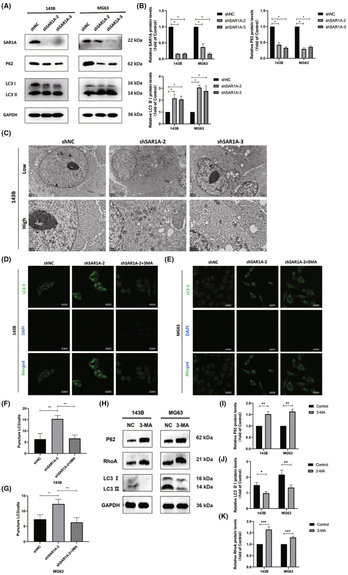
FIGURE 6.

Knocking down SAR1A in osteosarcoma cells induces autophagic activity. (A, B) Autophagy‐associated protein levels in MG63 and 143B cells were assessed by western blotting, with GAPDH being used for normalization. (C) Representative transmission electron microscopy images highlighting ultrastructural changes consistent with the induction of autophagic activity in 143B cells following shSAR1A or normal control (shNC) transfection. (D–G) Punctate LC3II fluorescence was evident in 143B and MG63 cells following SAR1A knockdown in cells that were or were not treated with 3‐methyladenine (3‐MA; 5 mmol/L) for 12 h. (H–K) RhoA expression was decreased in 143B and MG63 cells after 12 h treatment with 3‐MA (5 mmol/L). Analyses were repeated three times. * p <0.05, ** p <0.01, *** p <0.001 (mean ± SD).
